
Mouse Secondary Antibodies

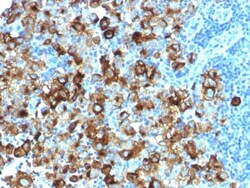
Biotium gp100 / Melanosome / PMEL17 / SILV (Melanoma Marker) (PMEL/1825R), 1mg/mL
Cytotoxic T lymphocytes (CTLs) recognize melanoma-associated antigens, which belong to three main groups. These groups include tumor-associated testis-specific antigens, melanocyte differentiation antigens and mutated or aberrantly expressed antigens, which are routinely used as markers to identify melanomas based on their binding to specific monoclonal antibodies. gp100, also designated ME20-M, ME20-S and PMEL 17, is classified as a melanocyte differentiation antigen and is expressed at low levels in normal cell lines and tissues, but is upregulated in melanocytes. gp100 is a highly glycosylated protein. It is also the product of proteolytic cleavage, which results in a secreted protein.Primary antibodies are available purified, or with a selection of fluorescent CF Dyes and other labels. CF Dyes offer exceptional brightness and photostability. Note: Conjugates of blue fluorescent dyes like CF405S and CF405M are not recommended for detecting low abundance targets, becau


Biotium Mucin 5AC (MUC5AC)(CLH2), CF740 conjugate, 0.1mg/mL
Mucin 5AC glycoprotein (MUC5AC) is a HMW glycoprotein belonging to the superfamily of mucins. Mucins are produced by epithelial cells and can be divided into two families; secretory mucins and membrane bound mucins. MUC5AC is a mucus-forming, secreted mucin that is found in normal gastric and tracheo-bronchial mucosa, but absent from normal colon. MUC5AC expression is present in primary ovarian mucinous cancer but usually absent in colorectal adenocarcinoma, thus showing an expression pattern opposite to MUC2. Together with a panel of antibodies, Anti-MUC5AC may be useful for differential identification of primary mucinous ovarian tumors from colon adenocarcinoma metastatic to the ovary. MUC5AC antibodies may also be useful for identification of intestinal metaplasia as well as in the identification of pancreatic carcinoma and pre-cancerous changes vs. normal pancreas.Primary antibodies are available purified, or with a selection of fluorescent CF Dyes and other labels.
Biotium Cytokeratin, HMW(KRTH/1076), Biotin conjugate, 0.1mg/mL
This MAb recognizes basic (Type II or HMW) cytokeratins, which include 67 kDa (CK1); 64 kDa (CK3); 59 kDa (CK4); 58 kDa (CK5); 56 kDa (CK6); 52 kDa (CK8). Twenty human keratins are resolved with two-dimensional gel electrophoresis into acidic (pI 6.0) subfamilies. The acidic keratins have molecular weights (MW) of 56.5, 55, 51, 50, 50 , 48, 46, 45, and 40 kDa. Many studies have shown the usefulness of keratins as markers in cancer research and tumor diagnosis.Primary antibodies are available purified, or with a selection of fluorescent CF Dyes and other labels. CF Dyes offer exceptional brightness and photostability. Note: Conjugates of blue fluorescent dyes like CF405S and CF405M are not recommended for detecting low abundance targets, because blue dyes have lower fluorescence and can give higher non-specific background than other dye colors.
Biotium NKX2.2 (Neuroendocrine & Ewing s Sarcoma Marker) (NX2/1523), Biotin conjugate, 0.1mg/mL
Expression of NKX2.2 has been found in neuroendocrine tumors of the gut, making it a potential marker for the study of gastrointestinal neuroendocrine tumors. More recently, NKX2.2 protein was identified as a target of EWS-FLI-1, the fusion protein specific to Ewing sarcoma, and was shown to be differentially upregulated in Ewing sarcoma on the basis of array-based gene expression analysis. It acts as a valuable marker for Ewing sarcoma, with a sensitivity of 93% and a specificity of 89%, and aids in the differential diagnosis of small round cell tumors.Primary antibodies are available purified, or with a selection of fluorescent CF Dyes and other labels. CF Dyes offer exceptional brightness and photostability. Note: Conjugates of blue fluorescent dyes like CF405S and CF405M are not recommended for detecting low abundance targets, because blue dyes have lower fluorescence and can give higher non-specific background than other dye colors.
Biotium BOB.1 (B-Cell Marker) (BOB1/2425), 0.2mg/mL
BOB-1 is a transcriptional coactivator that specifically associates with either POU2F1/OCT1 or POU2F2/OCT2, boosting the promoter activity of those complexes. It is essential for the response of B-cells to antigens, required for the formation of germinal centers, and regilates IL-6 expression. BOB-1 expression in a variety of established B-cell lines, representing different stages of B-cell development, has suggested a constitutive, B-cell-specific expression pattern. LP cells in nodular lymphocyte predominant Hodgkin lymphoma, because they are germinal center-derived, are consistently immuno-positive for BOB-1. Conversely, only some cases of classical Hodgkin lymphoma show BOB-1 immuno-reactivity within the Hodgkin and Reed-Sternberg cells. Expression of BOB-1 has been reported in follicular center cell lymphoma, diffuse large B-cell lymphoma and some cases of acute myeloid leukemia. B-CLL, marginal zone lymphoma, and mantle cell lymphoma may show weak to moderate immunoreactivit
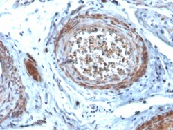
Biotium StAR (Steroidogenic Acute Regulator) (Leydig Cell Marker)(STAR/2077), CF568 conjugate, 0.1mg/mL
Steroidogenic Acute Regulatory Protein (STAR) controls the rate-limiting step of steroidegenesis by translocating cholesterol from the outer mitochondrial membrane to the inner membrane where it is later cleaved to pregnenolone. It is primarily present in steroid-producing cells, including Leydig cells in the testis, theca cells and luteal cells in the ovary and adrenal cells in the adrenal cortex. Due to low levels of pregnenolone, seminomas and Leydig cell tumors display no specific STAR staining. Therefore, STAR antibody may assist in differentiating sex cord stromal tumors (SCST), seminomas and embryonal carcinomas. Primary antibodies are available purified, or with a selection of fluorescent CF Dyes and other labels. CF Dyes offer exceptional brightness and photostability. Note: Conjugates of blue fluorescent dyes like CF405S and CF405M are not recommended for detecting low abundance targets, because blue dyes have lower fluorescence and can give higher non-specific back
Biotium MECA-79(MECA-79), 0.2mg/mL
MECA-79 recognizes a carbohydrate epitope shared with a group of sulfation decorated sialomucins, including sulfated ligands for CD62L (CD34, GlyCAM-1, Sgp200, and a subset of MAdCAM-1). This set of antigens has been referred to as peripheral node addressin (PNAd) with the molecular mass 50-250 kD. It has been identified that GlcNAc-6-SO4 sulfation contributes to MECA-79 binding and the core 1 beta1, 3-N-acetylglucosaminyltransferase is required for the generation of the MECA-79 epitope. MECA-79 is expressed on high endothelial venules (HEV) of lymphoid tissues, chronic inflamed tissues and rheumatoid synovia. The interaction of PNAd with CD62L receptor is involved in tethering and rolling of lymphocytes along HEV in lymphoid tissues. MECA-79 antibody reacts with PNAd from mouse, human and many other species and blocks L-selectin-dependent lymphocyte adhesion in vitro and in vivo. Primary antibodies are available purified, or with a selection of fluorescent CF Dyes and other
Biotium BOB.1 (B-Cell Marker) (BOB1/2423), CF488A conjugate, 0.1mg/mL
BOB-1 is a transcriptional coactivator that specifically associates with either POU2F1/OCT1 or POU2F2/OCT2, boosting the promoter activity of those complexes. It is essential for the response of B-cells to antigens, required for the formation of germinal centers, and regilates IL-6 expression. BOB-1 expression in a variety of established B-cell lines, representing different stages of B-cell development, has suggested a constitutive, B-cell-specific expression pattern. LP cells in nodular lymphocyte predominant Hodgkin lymphoma, because they are germinal center-derived, are consistently immuno-positive for BOB-1. Conversely, only some cases of classical Hodgkin lymphoma show BOB-1 immuno-reactivity within the Hodgkin and Reed-Sternberg cells. Expression of BOB-1 has been reported in follicular center cell lymphoma, diffuse large B-cell lymphoma and some cases of acute myeloid leukemia. B-CLL, marginal zone lymphoma, and mantle cell lymphoma may show weak to moderate immunoreactivit
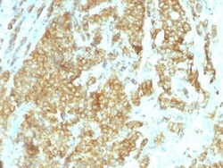
Biotium Glucose Regulated Protein 94 (GRP94)(HSP90B1/1192), Biotin conjugate, 0.1mg/mL
Recognizes a protein of 94 kDa, which is identified as the glucose-regulated protein 94 (grp94) and also tumor rejection antigen (gp96). Grp94 shows a high degree of sequence homology with the heat shock protein 90 (hsp90). This MAb is highly specific to grp94 and shows minimal cross-reaction with other members of the HSP90 family. Grp s are a class of proteins unresponsive to heat shock and are induced by glucose deprivation. Grp94 has been briefly studied as a prognostic factor in breast cancer.Primary antibodies are available purified, or with a selection of fluorescent CF Dyes and other labels. CF Dyes offer exceptional brightness and photostability. Note: Conjugates of blue fluorescent dyes like CF405S and CF405M are not recommended for detecting low abundance targets, because blue dyes have lower fluorescence and can give higher non-specific background than other dye colors.
Biotium Cytokeratin 19(KRT19/799 + KRT19/800), CF488A conjugate, 0.1mg/mL
Recognizes a protein of 40 kDa, identified as cytokeratin-19 (CK19), which is expressed in sweat gland, mammary gland ductal and secretory cells, bile ducts, gastrointestinal tract, bladder urothelium, oral epithelia, esophagus, and ectocervical epithelium. Anti-CK19 reacts with a wide variety of epithelial malignancies including adenocarcinomas of the colon, stomach, pancreas, biliary tract, liver, and breast. Perhaps the most useful application is the identification of thyroid carcinoma of the papillary type, although 50%-60% of follicular carcinomas are also labeled. Anti-CK19 is a useful marker for detection of tumor cells in lymph nodes, peripheral blood, bone marrow and breast cancer.Primary antibodies are available purified, or with a selection of fluorescent CF Dyes and other labels. CF Dyes offer exceptional brightness and photostability. Note: Conjugates of blue fluorescent dyes like CF405S and CF405M are not recommended for detecting low abundance targets, because
Biotium Mucin 5AC (MUC5AC)(58M1), 0.2mg/mL
This MAb recognizes the peptide core of gastric mucin M1 (recently identified as Mucin 5AC). Its epitope is destroyed by beta-mercaptoethanol but not by periodate treatment. This mucin is present in primary ovarian mucinous cancer but usually absent in colorectal adenocarcinoma, thus showing an expression pattern opposite to MUC2. Together with a panel of antibodies, Anti-MUC5AC may be useful for differential identification of primary mucinous ovarian tumors from colon adenocarcinoma metastatic to the ovary. MUC5AC antibodies may also be useful for identification of intestinal metaplasia as well as in the identification of pancreatic carcinoma and pre-cancerous changes vs. normal pancreas.Primary antibodies are available purified, or with a selection of fluorescent CF Dyes and other labels. CF Dyes offer exceptional brightness and photostability. Note: Conjugates of blue fluorescent dyes like CF405S and CF405M are not recommended for detecting low abundance targets, becau
Biotium CD99 / MIC2 (Ewing s Sarcoma Marker) (MIC2/1495R), 0.2mg/mL
This antibody recognizes a sialoglycoprotein of 27-32 kDa, identified as CD99, or MIC2 gene product, or E2 antigen. MIC2 gene is located in the pseudo-autosomal region of the human X and Y chromosome. MIC2 gene encodes two distinct proteins, which are produced by alternative splicing of the CD99 gene transcript and are identified as bands of 30 and 32 kDa (p30/32). Although its function is not fully understood, CD99 is implicated in various cellular processes including homotypic aggregation of T cells, upregulation of T cell receptor and MHS molecules, apoptosis of immature thymocytes and leukocyte diapedesis. CD99 is expressed on the cell membrane of some lymphocytes, cortical thymocytes, and granulosa cells of the ovary. Most pancreatic islet cells, Sertoli cells of the testis, and some endothelial cells express this antigen. Mature granulocytes express very little or no CD99. MIC2 is strongly expressed on Ewing s sarcoma cells and primitive peripheral neuroectodermal tumor

Biotium N-Cadherin / Cadherin-2 / CD325 (NCAD) (13A9), CF568 conjugate, 0.1mg/mL
This antibody recognizes a protein of ~140 kDa, identified as N-Cadherin (NCAD), also known as CD325. NCAD is a member of the Cadherin superfamily, and consists of five extracellular repeats, a transmembrane domain and a cytoplasmic domain. CD325 deficient mice die at day 10 of gestation and embryos display major heart defects and malformed neural tubes and somites. Consistent with this, CD325 has been implicated in several aspects of cardiac development including the precardiac mesoderm, establishment of left-right symmetry and cardiac looping morphogenesis. Furthermore, CD325 is normally involved in inducing cell cycle arrest and its expression is frequently deregulated in cancer cells. Studies have linked N-cadherin to cancer metastasis by showing the aggressive tumor cells had preferentially turned on N-cadherin as opposed to E- or P-cadherin.Primary antibodies are available purified, or with a selection of fluorescent CF Dyes and other labels. CF Dyes offer exceptional
Biotium Galectin-13 (GAL13) / Placental Protein 13 (PP13) (PP13/1165), 1mg/mL
This antibody recognizes a 32 kDa protein, which is identified as homodimer of galectin-13 (also known as PP13). Galectins are a family of soluble beta-galactoside-binding lectins that modulate cell-to-cell adhesion and cell-to-extracellular matrix (ECM) interactions and play a role in tumor progression, pre-mRNA splicing and apoptosis. One such member, galectin-13, is a 139 amino acid protein that contains one galectin domain. Possessing lysophospholipase activity, galectin-13 exists as a disulfide-linked homodimer. Galectin-13 is suggested to have a developmental role in the placenta and may display immunobiological activity in feto-maternal blood-spaces. Screening for galectin-13 in maternal serum during the first trimester of pregnancy may serve as a diagnostic marker to predict preterm pre-eclampsia.Primary antibodies are available purified, or with a selection of fluorescent CF Dyes and other labels. CF Dyes offer exceptional brightness and photostability. Note: Co
Biotium Cytokeratin 7(KRT7/1198), CF568 conjugate, 0.1mg/mL
This antibody recognizes an intermediate filament protein (IFP) of 55 kDa, which is identified as cytokeratin 7. This MAb is highly specific to cytokeratin 7 and shows no cross-reaction with other IFPs. Cytokeratin 7 is a basic cytokeratin, which is found in most glandular and transitional epithelia but not in the stratified squamous epithelia. Keratin 7 is expressed in the epithelial cells of ovary, lung, and breast but not of colon, prostate, or gastrointestinal tract. This MAb is highly useful in distinguishing ovarian carcinomas (keratin 7 ) from colon carcinomas (keratin 7-).Primary antibodies are available purified, or with a selection of fluorescent CF Dyes and other labels. CF Dyes offer exceptional brightness and photostability. Note: Conjugates of blue fluorescent dyes like CF405S and CF405M are not recommended for detecting low abundance targets, because blue dyes have lower fluorescence and can give higher non-specific background than other dye colors.